
It's Medical Monday: Minimally Invasive Aortic Surgery
- keyanazahiri
- Jul 14, 2025
- 2 min read
Welcome back to Medical Monday! Today, we will be continuing on our surgery theme of the month and covering minimally invasive aortic surgery. In the last few weeks, we’ve discussed some of the most common traditional open approaches to surgery on the heart and lungs via sternotomy and thoracotomy. Make sure to refer back to those posts for a refresher before we dive into the minimally invasive approach in this post.
Cardiac surgery is typically thought of as one of the most open and invasive forms of surgery, as large access is typically needed to repair issues with the aorta and heart that sit behind the sternum and ribs. However, recent medical advancements have allowed for some procedures to be performed in a more minimally invasive manner via smaller incisions for patients and avoidance of many of the risks of open surgery. When it comes to minimally invasive aortic surgery, Thoracic Endovascular Aortic Repair (TEVAR) is the most common approach. Sometimes, these procedures will be done in collaboration with our vascular surgery colleagues. In patients who are suitable candidates, TEVAR is often favored over open surgery due to the reduced risks of minimally invasive approaches and quicker recovery times. By now, we are all generally familiar with the basics of aortic anatomy, aortic aneurysms, and dissections. Feel free to refer back to our previous blog posts for a quick review.
TEVAR is most commonly used to repair aneurysms and dissections of the descending thoracic aorta, the segment of the aorta after the arch that runs down through your chest and towards your belly. Prior to receiving a TEVAR, patients receive a thorough workup to ensure they are a suitable candidate. A number of imaging tests will be used to first ensure there is enough space in your vessels for the catheters to reach the thoracic aorta and also assist with selecting the appropriate size and type of stent graft. In general, the basic steps of a TEVAR involves gaining access to the damaged segment of the thoracic aorta with a guide wire inserted through a small incision in your femoral artery, located in the upper thigh near the groin. Then, using fluoroscopy to allow for real time imaging visualization of the inside of vessels and other internal structures, a catheter is inserted over the guidewire with the stent graft that will be placed into the damaged area of the aorta. After confirming with imaging that the correct area of the aorta has been accessed, the stent graft is deployed, expanding to sit against the walls of the aorta and provide a stronger structure for blood to flow through.

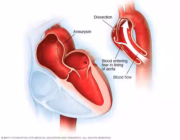
Above, you can see a graphic depicting the overall concept of a TEVAR and here you can view a brief video providing an overview and answering some common patient questions regarding TEVAR: https://www.youtube.com/watch?v=8TOIg0AghOQ. Not all types of aortic aneurysms are suitable for TEVAR, however, so it is important to consult your provider and medical team to determine if you are an appropriate candidate. As always, please let us know if you have any questions and feel free to refer to our previous blog posts for more information.
Best wishes,
Keyana Zahiri
Brown Medical Student - MS3
Sources:


Comments