
It's Medical Monday
- Samantha Colon
- Apr 1, 2024
- 2 min read

Updated: Jun 22, 2025
Hi everyone! My name is Samantha and I'm a medical student at the CUNY School of Medicine. I'll be taking over Medical Mondays and today I'm going to discuss some of the imaging techniques mentioned last week.
Let's delve into computed tomography (CT) imaging. CT scans are the gold standard for diagnosing aortic aneurysms and dissections.
Why is a CT scan done?
CT scans are frequently used for aortic aneurysms and dissection diagnoses due to their high resolution and ability to provide detailed images of the aorta and related structures. As we all know, aortic aneurysms and dissections are serious conditions that require prompt action and treatment to prevent complications such as rupture or organ damage. Physicians order CT scans to accurately assess the size, location, and extent of the abnormality.
How is a CT scan done?

A CT scan uses X-ray and computer technology to create detailed cross-sectional images. Contrast dye may be given to visualize the aorta and associated blood vessels better. Patients lay on a bed that slides through the CT scanner, which then captures images as it rotates around them. This part usually only takes 10 minutes, but the entire process can take up to 60 minutes. Once all of the images are taken, they are reconstructed to create a three-dimensional picture that healthcare professionals can analyze.
Where is a CT scan done?
This imaging technique can be performed at hospitals, imaging centers, or outpatient clinics. Some centers may not have an available CT scanner on-site, as resources vary between facilities. In emergent situations, like an aortic dissection, imaging may be conducted in the emergency department.
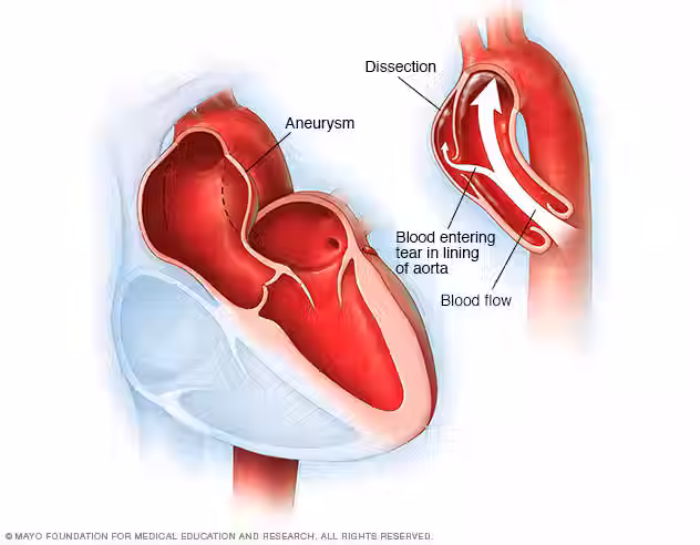
What does an aortic aneurysm look like on CT?

As you can see from the picture above, an aneurysm looks like a balloon-like bulge coming from the aorta. The size and shape can differ depending on the underlying cause and location. However, all aneurysms usually appear as a well-defined dilation.
What does an aortic dissection look like on CT?

In this image, the arrow is pointing to a tear in the aortic wall. Now since there are two lumens, blood can flow through both, which is not supposed to happen! A CT image with contrast can show the path of blood flow and can be used to assess the extent and severity of the dissection.
Are there risks to a CT scan?
CT scans use X-rays, as mentioned before, which use ionizing radiation. Radiation can damage your DNA and increase your risk of developing cancer. However, current research shows the chance of this happening is very low- about 1 in 2,000. The benefits of a CT scan, especially if being used in an emergent aortic dissection situation, overly outweigh any potential adverse effect. Remember to talk to your doctor about the specific risks and benefits of any procedure.
That wraps up this week's Medical Monday. I hope you were able to learn a thing or two about CT scans! Join me in two weeks to learn about MRIs and if it's better, worse, or just as good as CT scans.
Have a great week, and always Think Aorta.
Samantha


wow this helps a lot knowing what to expect for my ct scan and how it all works thanks samantha
Excellent information.
Thank You!